
A correct diagnosis is important. Not only to get the best help possible, but also because otherwise the public image of Visual Snow (VS) and Visual Snow Syndrome (VSS) is misrepresented with contributions from people who think they have it. To receive a correct diagnosis, it is important that if you have VS(S) symptoms to see a doctor who will rule out other conditions that may overlap with VS(S) in terms of symptoms.
In this research paper (link to: https://thejcn.com/DOIx.php?id=10.3988/jcn.2020.16.4.646) neuro-ophthalmologists recommend ruling out retinal and neurological diseases as possible causes of VS(S).
And in this research paper (link to: https://link.springer.com/article/10.1007/s11910-018-0854-2) doctors are advised to rule out possibly other underlying diseases/conditions when receiving a patient with VS(S) symptoms.
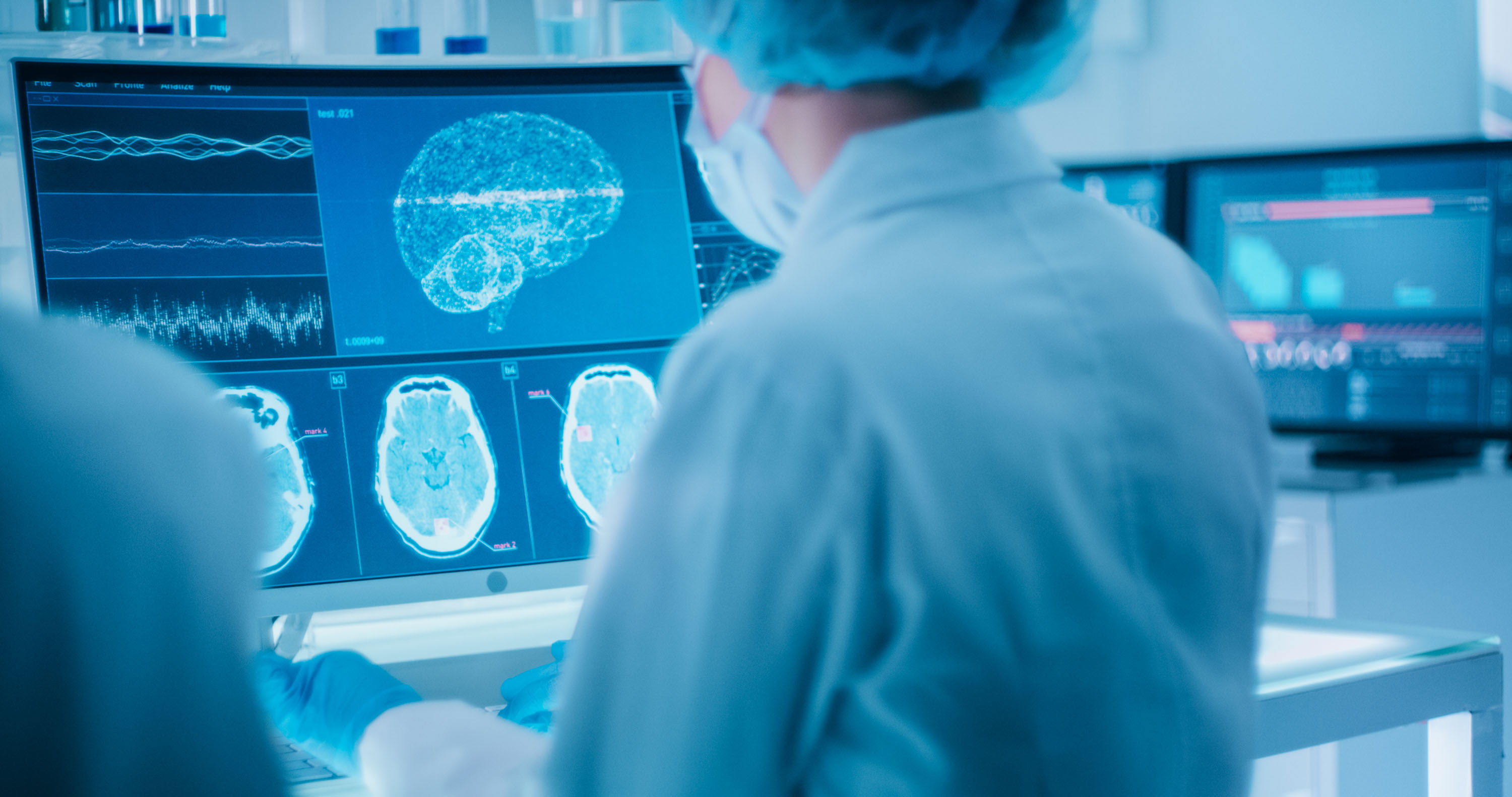
It is important to be examined by both a neurologist and an optometrist. First of all, this is a neurological disorder and also the main symptoms express themselves visually. An MRI scan, EEG and VEP test are important tests that should be used to get a better picture. Although a typical MRI scan will usually not be abnormal, it is important to exclude other conditions. A PET-/CT-scan has shown abnormalities in recent research (link: https://academic.oup.com/brain/article/143/4/1106/5811372), however, such a scan can only be interpreted by a neurologist who is familiar with VS(S). VS(S) patients showed hypermetabolism and cortical volume increase in the extrastriate visual cortex at the junction of the right lingual and fusiform gyrus. There was hypometabolism in the right superior temporal gyrus and the left inferior parietal lobule. Patients had increases in gray matter volume in the temporal and limbic lobes and decreases in the superior temporal gyrus. The corresponding structural and functional changes highlight the relevance of the visual association cortex to VSS. This gives the clinical impression that the disorder extends beyond the visual system.
A VEP test shows no abnormalities in the classical sense, however, if VS(S) is present, increased latency is often seen when paying attention to N145. And this is typical for VS(S). This points in the direction of impaired processing in the secondary visual areas and hyperactivation in the primary visual cortex. VS(S) patients have subtle, significant neuroanatomical differences in the major visual and lateral cerebellar areas, which may partially explain the pathophysiological basis of the disorder.
Disclaimer:
This information is for informational purposes only and should not be used as a substitute for medical advice from a licensed professional.